

How To Assess Your Dog’s Cancer Risk
Prevention and early detection can save your dog’s life—here’s what to look for
Knowing your dog’s cancer risk is absolutely key to cancer prevention. By identifying dogs at an increased risk of cancer development, we can watch for warning signs, allowing for diagnosis at a stage when intervention will be most successful.
Assessing an individual dog’s cancer risk is accomplished by taking into account conventional risk factors—age, breed, gender, family history, carcinogen exposure—to identify dogs generally at risk of developing certain cancers. From this information, specific screening and surveillance programs along with recommendations for cancer prevention can be implemented. Here’s how to assess your dog’s risk factor, along with cancer warning signs to watch for.
Cancer Screening: What To Watch For
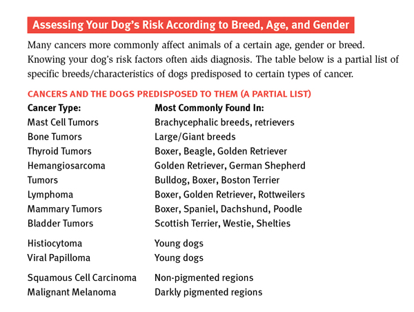
Unfortunately, it is well known that certain breeds are prone to develop cancer. Worse still, because cancer is a common disorder of older dogs and cats, all pets beyond the age of seven or eight years of age should be considered “at risk” for cancer. Annual physical exams, screening laboratory bloodwork, and urinalysis are recommended for dogs over seven years of age. In addition, the use of cutaneous maps to chart the location, size, and diagnosis of all skin masses will help to determine rapid changes in growth or any new masses to be concerned about. Ask your vet to chart all skin masses.
Owners must also take responsibility for prevention of cancer. Early neutering/spaying of male and female dogs is the best example of prevention of testicular and mammary cancer. Certain breeds of dogs with long noses, such as the Collies or Shepherds, are at increased risk of developing upper respiratory disease as well as cancer when exposed to second-hand smoke and should be removed from a passive smoke environment. (Note that second-hand smoke is bad for all dogs but particularly terrible for those already predisposed to upper respiratory cancer.) Likewise, frequent use of coal or kerosene as indoor heating sources may increase the risk of nasal cancers. Observation of bowel or urinary habits should be consistently performed. Any concern about straining or blood should be checked out by your vet. Owners should also be able to accurately evaluate mammary glands, peripheral lymph nodes, oral cavity structures, examine interdigital spaces, and external ear canals. Ask your vet to show you what to look for.
What To Do If You Find Something
Noting the onset and duration of the mass, its growth rate, and prior treatment will help determine a course of action. Any previous tumors diagnosed in the patient should be identified as to type, completeness of treatment and follow up. The history of when a female dog was spayed or the reproductive history should be detailed.
Physical Examination: The goal of an examination is to identify any other issues or diseases that may limit cancer treatment options and to define the extent of the tumor burden. A topographic map of the patients’ body is useful for future comparison and for identifying benign skin lesions such as lipoma and sebaceous adenomas. Physical measurements of any superficial lesion(s) using calipers is useful to document changes in size of superficial masses, most of which are not malignant. It is essential to estimate the invasiveness of a tumor and the degree of attachment to underlying tissue in order to adequately plan the surgical biopsy and/or resection of a lesion. Regional lymph nodes must be evaluated for size, consistency, and fixation to adjacent tissues. Physical exam findings will also help in selection of ancillary diagnostic procedures necessary to define tumor extent (specific imaging techniques, type of biopsy, endoscopy, etc.)
Diagnostic Evaluation—Clinical Evaluation: After a thorough physical examination, a screening laboratory evaluation should follow. This generally includes a complete blood cell count, serum biochemistry panel, and urinalysis. Other diagnostic tests are performed as indicated. Survey radiographs are suggested to detect metastasis, determine potential bone involvement or evaluate orthopedic soundness prior to amputation or limb-sparing in dogs with osteosarcoma, localize oral or nasal masses, etc. The next step is CAT scans and MRIs, both of which are routinely available and are able to define the invasive characteristics of deep-seated tumors much more clearly than survey radiographs. They may be particularly helpful when planning involved surgical procedures. Ultrasonography can be used to determine the proximity of a tumor to large blood vessels, to determine the cystic nature of masses, to evaluate possible intra-abdominal metastases to lymph nodes or organs, and to assess the initial and post-treatment tumor volume.
 Cytologic examinations (examination of tissue samples) from fine needle aspiration biopsies of accessible tumors and regional lymph nodes are important diagnostic procedures. Fine needle aspiration can be accomplished on any accessible mass. Often, a rapid, inexpensive diagnosis can be made for certain tumor types (lipomas, sebaceous adenomas, lymphoma and mast cell tumors). Treatment decisions should be considered on a cytologic diagnosis only when a definitive diagnosis can be made, such as with lymphoma or mast cell tumors.
Tumor Biopsy: Many techniques are available for tissue biopsy. The method selected should safely and simply procure adequate tissue samples to provide an accurate diagnosis without compromising treatment. Biopsies can be excisional (complete removal of a tumor that is small and not attached to underlying tissues) or nonexcisional (removal of only a portion of the tumor). Nonexcisional techniques include: a) a fine-needle aspirate, tissue impression smears or fluid analysis or b) tissue samples procured from a surgical incision or punch biopsy.
Even if a cancer diagnosis is possible with a fine needle aspiration, a tissue biopsy may be indicated to establish prognosis and guide treatment planning. For example, the histologic grade of soft tissue sarcomas and mast cell tumors are prognostic factors that can be helpful in treatment planning. Biopsy results may suggest the degree of surgical resection necessary for definitive removal and a clean margin or indicate that additional types of therapy may be beneficial.
Each dog and pet owner have unique circumstances that must be considered. Your veterinarian should provide thorough, accurate, and unbiased options for management based on an understanding of the patients’ general health, specific tumor type, the likelihood of response to treatment, maintenance of good quality of life, and emotional support for owners. Each step of the diagnosis and therapy plan can and should be considered carefully. Many new techniques, procedures and management information are available. Consultation by a specialist is one of the best investments an owner with a pet that has cancer can make.

Join the newsletter and never miss out on dog content again!
"*" indicates required fields
By clicking the arrow, you agree to our web Terms of Use and Privacy & Cookie Policy. Easy unsubscribe links are provided in every email.